Pump Station Slides
Biology Lab • 45-Minute Mission
Cardiovascular System
Vital Signs & Anatomical Pathways
PUMP STATION:
The Anatomy of Human Blood Circulation
Trace the 60,000-mile highway inside your body, unlock the four muscular chambers, and measure your own mechanical pump in action.
1
4 Chambers & Valves
2
Pulmonary vs Systemic
3
Pulse & Output Lab
The Engine: Facts Behind the Fist
Stage 1 • Hook
Daily Rhythms
100,000
Beats every single day without taking even a 2-second break. Over 2.5 billion beats in an average lifetime!
Fluid Volume
2,000 Gal
Pumps approximately 7,500 liters of blood each day—enough to fill an entire backyard swimming pool every week.
Vessel Pipeline
60,000 Mi
Combined length of all your arteries, capillaries, and veins—long enough to wrap around the Earth more than twice.
Cardiac Muscle (Myocardium): Unlike skeletal muscles that fatigue quickly, cardiac muscle fibers are interconnected by intercalated discs that contract rhythmically and resist exhaustion.
The 4 Chambers: Mind the Anatomical Reverse!
Key Rule: Patient's Right = Your Left!
RIGHT SIDE (Deoxygenated) Heading to Lungs
1. Right Atrium
Receives oxygen-depleted blood returning from body via Superior & Inferior Vena Cava.
2. Right Ventricle
Muscular lower pump that forces blood into Pulmonary Artery toward the lungs.
LEFT SIDE (Oxygenated) Heading to Body
3. Left Atrium
Receives oxygen-rich blood returning fresh from the lungs via Pulmonary Veins.
4. Left Ventricle (The Heavy Lifter)
Thickest muscle wall; builds huge pressure to launch blood into Aorta for whole body.
Quick Rule of Thumb: ATRIA = Always Receive • VENTRICLES = Always Pump Septum divides left and right
One-Way Valves: Making the “Lub-Dub”
Fluid Dynamics
1st Sound: “LUB”
Caused by the snapping shut of the Atrioventricular (AV) Valves:
- Tricuspid Valve (Right atrium → Right ventricle)
- Bicuspid / Mitral Valve (Left atrium → Left ventricle)
Function: Prevents blood from regurgitating backwards into the atria during ventricular squeeze.
2nd Sound: “DUB”
Caused by the snapping shut of the Semilunar (SL) Valves:
- Pulmonary Valve (Right ventricle → Pulmonary trunk)
- Aortic Valve (Left ventricle → Aorta)
Function: Closes as ventricles relax to stop pressurized arterial blood from draining back down.
Clinical Connection: A heart murmur is caused by turbulent backflow when a valve fails to seal tightly!
The Dual Highway: Two Interconnected Loops
Figure-8 Circuit
Loop 1
Pulmonary Circuit
Mission: Gas Exchange in the Lungs
Right Ventricle
↓ Pulmonary Arteries (O2-poor)
↓ Lung Capillaries (drops CO2, picks up O2)
↓ Pulmonary Veins (O2-rich)
↓ Left Atrium
Unique Exception: Pulmonary arteries are the only arteries carrying deoxygenated blood!
Loop 2
Systemic Circuit
Mission: Fuel Every Cell in the Body
Left Ventricle
↓ Aorta & Major Arteries
↓ Systemic Capillaries (delivers O2 to cells)
↓ Vena Cava (Veins returning O2-poor)
↓ Right Atrium
High Pressure System: Left ventricle is 3× thicker because it pushes blood all the way to toes and brain!
Arteries = Away from heart • Veins = Visit back to heart • Capillaries = Crossover delivery
Vessel Trio & The Big Myth
Biomedical Truth Check
Arteries
Thick, elastic muscular walls that withstand explosive systolic pressure surges.
Pulse point: Radial & Carotid
Capillaries
Microscopic single-cell layer thin. Red blood cells squeeze through in single file!
Diffusion of O2, CO2 & nutrients
Veins
Lower pressure, wider lumen, and equipped with flap valves to prevent gravity pooling.
Skeletal muscle pump helps flow
MYTH BUSTED: Deoxygenated Blood Is NOT Blue!
Blood is never blue inside human vessels. Oxygenated blood is bright scarlet red; deoxygenated blood is dark burgundy/brick red. Veins look bluish through skin purely because of light wavelengths scattering through skin layers!
Lab Protocol: Pulse & Circuit Investigation
18 Minutes • Team of 2
Part 1: Circuit Trace
Follow a single red blood cell entering from the Vena Cava. Number the sequence 1 to 10 through chambers, valves, lungs, aorta, and tissues.
Color code: BLUE = O2-Poor | RED = O2-Rich
Part 2: Heart Rate & Output
Locate radial pulse (wrist). Count beats for 15 sec × 4 to find Resting BPM. Test light stress/elevation response and calculate Cardiac Output!
Cardiac Output = Heart Rate (BPM) × Stroke Volume (70 mL)
Materials Ready: Lab Handout • Timer / Stopwatch • Blue & Red Colored Pencils Let's Begin!
Heart Circuit Lab Worksheet
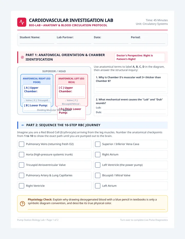
Cardiovascular Investigation Lab
BIO-LAB • ANATOMY & BLOOD CIRCULATION PROTOCOL
Time: 45 Minutes
Unit: Circulatory Systems
Student Name:
Lab Partner:
Date:
Period:
Part 1: Anatomical Orientation & Chamber Identification
Doctor's Perspective: Right is Patient's Right!
Superior / Head
Anatomical Right (O2-Poor)
[ A ] Upper Chamber:
↓ Valve [ E ]: Tricuspid ↓
[ B ] Lower Pump:
Anatomical Left (O2-Rich)
[ C ] Upper Chamber:
↓ Valve [ F ]: Bicuspid/Mitral ↓
[ D ] Thick Lower Pump:
Dividing Muscular Wall: Septum
Use anatomical terms to label A, B, C, D in the diagram, then answer the structural inquiry:
1. Why is Chamber D's muscular wall 3× thicker than Chamber B?
2. What mechanical event causes the "Lub" and "Dub" sounds?
Lub:
Dub:
Part 2: Sequence the 10-Step RBC Journey
Imagine you are a Red Blood Cell (Erythrocyte) arriving from the leg muscles. Number the anatomical checkpoints from 1 to 10 to show the exact path until you are pumped out to the brain.
Pulmonary Veins (returning fresh O2)
Superior / Inferior Vena Cava
Aorta (high-pressure systemic trunk)
Right Atrium
Tricuspid Atrioventricular Valve
Left Ventricle (the power pump)
Pulmonary Artery & Lung Capillaries
Bicuspid / Mitral Valve
Right Ventricle
Left Atrium
Physiology Check: Explain why drawing deoxygenated blood with a blue pencil in textbooks is only a symbolic diagram convention, and describe its true physical color.
Pump Station Biology Lab • Page 1 of 2 Turn over to complete Live Pulse Diagnostics
Part 3: Live Pulse Diagnostics & Physiological Response
Radial Pulse Investigation
Place two fingers on the thumb side of your wrist (radial artery). Do not use your thumb (it has its own pulse). Count beats for 15 seconds, multiply by 4 to determine Beats Per Minute (BPM).
| Condition | 15-Sec Count (You) | Heart Rate (BPM = Count × 4) | Partner's BPM |
|---|
| 1. Baseline Resting (Sitting quietly 2 min) |
Pump Station Teacher Guide
Pump Station • Teacher Facilitation Guide
45-MINUTE INSTRUCTIONAL PACING • LAB PROTOCOL • MASTER KEY
Biology / Life Science
Grades 8–12
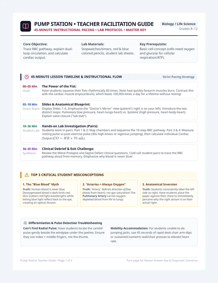
Core Objective: Trace RBC pathway, explain dual-loop circulation, and calculate cardiac output.
Lab Materials: Stopwatches/timers, red & blue colored pencils, student lab sheets.
Key Prerequisite: Basic cell concept (cells need oxygen and glucose for cellular respiration/ATP).
45-Minute Lesson Timeline & Instructional Flow
Strict Pacing Strategy
00–05 Min
Hook
The Power of the Fist:
Have students squeeze their fists rhythmically 60 times. Note how quickly forearm muscles burn. Contrast this with the cardiac muscle (myocardium), which beats 100,000 times a day for a lifetime without resting!
05–18 Min
Direct Teach
Slides & Anatomical Blueprint:
Display Slides 1–6. Emphasize the "Doctor's Mirror" view (patient's right is on your left). Introduce the two distinct loops: Pulmonary (low pressure, heart-lungs-heart) vs. Systemic (high pressure, heart-body-heart). Explain valve closure ("lub-dub").
18–36 Min
Student Lab
Hands-on Lab Investigation (Pairs):
Students work in pairs. Part 1 & 2: Map chambers and sequence the 10-step RBC pathway. Part 3 & 4: Measure resting pulse vs post-exercise pulse (30s high-knees or vigorous jumping), then calculate individual Cardiac Output (\(CO = HR \times 70\text{ mL}\)).
36–45 Min
Synthesis
Clinical Debrief & Exit Challenge:
Review the Mitral Prolapse and Septal Defect clinical questions. Cold-call student pairs to trace the RBC pathway aloud from memory. Emphasize why blood is never blue!
Top 3 Critical Student Misconceptions
1. The "Blue Blood" Myth
Truth: Human blood is never blue. Deoxygenated blood is dark brick-red. Skin scatters red light wavelengths while letting blue light reflect back to the eye, creating an optical illusion.
2. "Arteries = Always Oxygen"
Truth: "Artery" defines direction of flow (Away from heart), not gas saturation! The Pulmonary Artery carries oxygen-depleted blood from RV to lungs.
3. Anatomical Inversion
Truth: Students consistently label the left side as right. Have students place the paper against their chest to immediately perceive why the right atrium is on their actual right.