DNA Architecture Slides
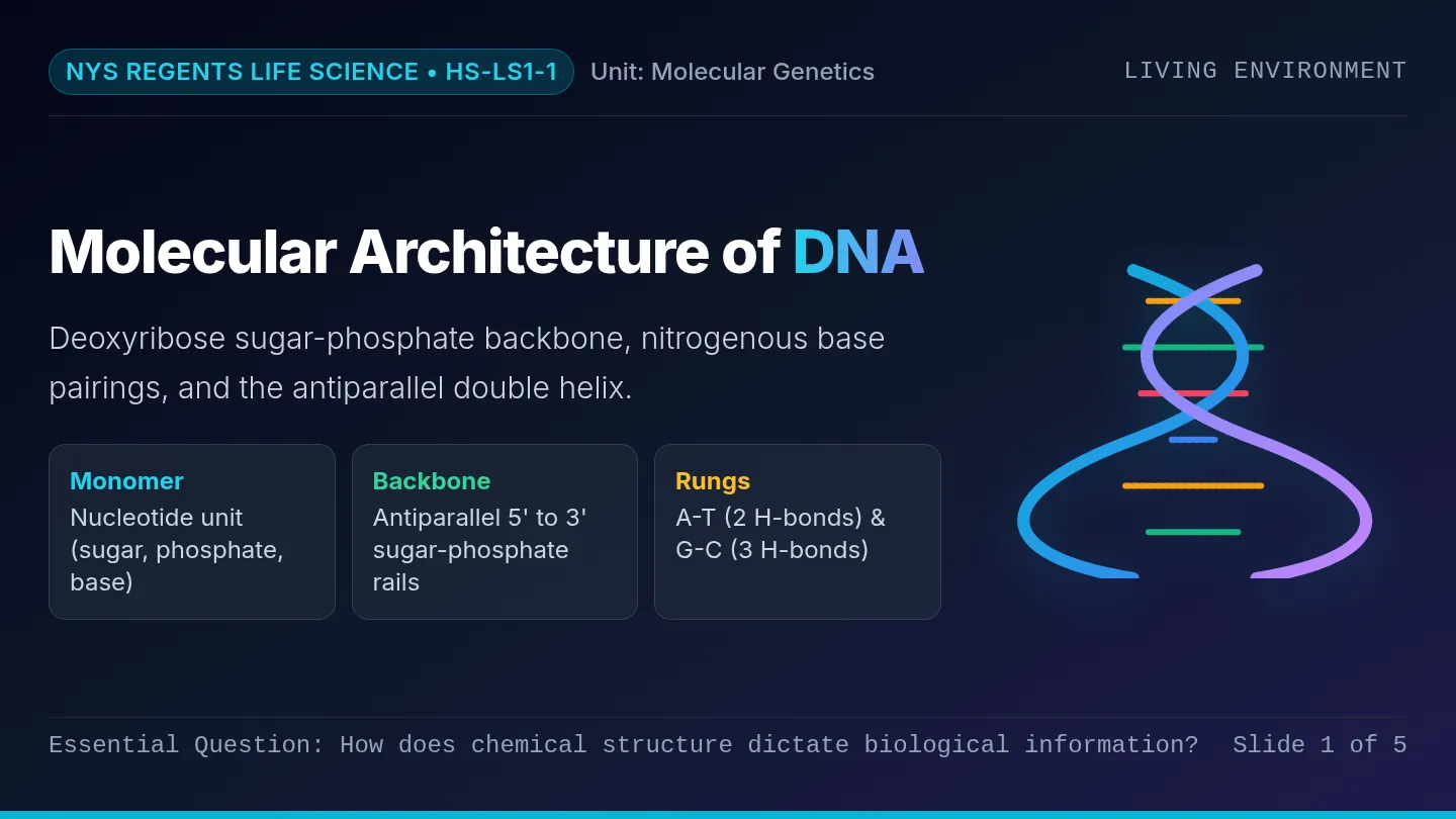
NYS Regents Life Science • HS-LS1-1 Unit: Molecular Genetics
Living Environment
Molecular Architecture of DNA
Deoxyribose sugar-phosphate backbone, nitrogenous base pairings, and the antiparallel double helix.
Monomer
Nucleotide unit (sugar, phosphate, base)
Backbone
Antiparallel 5' to 3' sugar-phosphate rails
Rungs
A-T (2 H-bonds) & G-C (3 H-bonds)
Essential Question: How does chemical structure dictate biological information? Slide 1 of 5
Part 1 • The Monomer
Anatomy of a Single Nucleotide
Key Regents Concept: Repeating Subunit
PO₄³⁻ Phosphate 5' C O Deoxyribose (5-Carbon Sugar) 1' 2' 3' (-OH) 4' Nitrogenous Base (A, T, C, or G) Phosphate Group 5-Carbon Deoxyribose Sugar Contains Genetic Code (N-Base)
1. Phosphate Group
Carries a negative charge (\(\text{PO}_4^{3-}\)); gives DNA its overall acidic and polar character.
2. Deoxyribose (5-Carbon Sugar)
Lacks oxygen at 2' position ("deoxy"). 5' carbon binds phosphate; 3' carbon binds the next nucleotide.
3. Nitrogenous Base
Attached to the 1' carbon. The variable component whose precise sequence encodes hereditary traits.
Regents Tip: All 3 parts covalently link to form ONE single nucleotide unit. Slide 2 of 5
Part 2 • Core Schematic Diagram
The Double Helix: Sugar-Phosphate Backbone & Base Pairs
Left Strand: 5' → 3' Right Strand: 3' → 5' (Antiparallel)
5' to 3' Direction 5' to 3' (Pointing Up) 5' End (Phosphate) 3' End (-OH Hydroxyl) 3' End (-OH Hydroxyl) 5' End (Phosphate) P D Adenine (A) 2 H-Bonds Thymine (T) D P P D Guanine (G) 3 H-Bonds Cytosine (C) D P OH Covalent Bond (Strong backbone) Hydrogen Bonds Weak (Easily Unzipped)
P = Phosphate
D = Deoxyribose
Sugar & phosphate form the outer structural rails.
Antiparallel Run
Strands point in opposite directions. Notice the deoxyribose pentagons point opposite ways!
Double Helix
The ladder twists into a right-handed spiral in 3D cellular space.
Regents Key: Strong covalent rails prevent breakage; weak H-bonds allow unzipping during replication. Slide 3 of 5
Part 3 • Complementarity & Bonding
Chargaff's Rules & Bond Thermodynamics
Purines (2 Rings) Pair with Pyrimidines (1 Ring)
Complementary Base Pairs (Chargaff)
A Adenine
== 2 H-Bonds == Weak Attractions
Thymine T
G Guanine
≡ 3 H-Bonds ≡ Slightly Stronger
Cytosine C
Regents Rule: In double-stranded DNA, %A = %T and %G = %C.
Covalent vs. Hydrogen Bonds
Covalent Rails
- • Very strong bonds
- • Sugar-to-phosphate
- • Keeps order intact
Hydrogen Rungs
- • Weak attractions
- • Between base pairs
- • Unzips for replication
Biological Advantage: Weak hydrogen bonds let helicase easily unzip DNA without destroying the backbone.
Standard Check: If a DNA molecule has 30% Adenine, what percentage is Cytosine? (Answer: 20%) Slide 4 of 5
Part 4 • Regents Synthesis
Regents Exam Question Analysis
Essential Life Science Mastery
Regents Practice Item 1 Structure Identification
A student models DNA using plastic pieces: pentagons, circles, and 4 colors of notched blocks. What does each piece represent?
• Pentagon: Deoxyribose (5-carbon sugar)
• Circle: Phosphate group (\(\text{PO}_4^{3-}\))
• 4 Notched Blocks: Nitrogen bases (A, T, C, G)
Regents Trap: Students often confuse the sugar with the phosphate or the nucleotide with the base.
Regents Practice Item 2 Replication & Bonding
During cell division, enzymes separate the two strands of DNA without breaking the strand itself. Which statement explains why?
Mastery Explanation: The two strands are held together by weak hydrogen bonds between base pairs, while the backbone is held by strong covalent bonds.
Therefore, helicase easily unzips the center without fragmenting the individual strands!
Next Step: Use the practice worksheet to label and assemble your own DNA nucleotide chain!
Exit Objective: Be able to sketch and label a 4-base-pair DNA fragment from memory. Slide 5 of 5
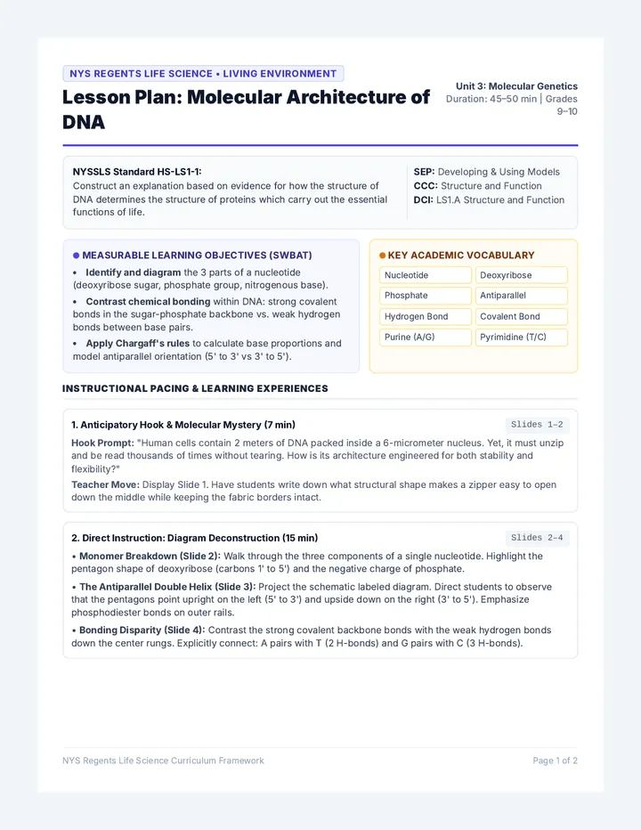
DNA Architecture Lesson Plan
NYS Regents Life Science • Living Environment
Lesson Plan: Molecular Architecture of DNA
Unit 3: Molecular Genetics
Duration: 45–50 min | Grades 9–10
NYSSLS Standard HS-LS1-1:
Construct an explanation based on evidence for how the structure of DNA determines the structure of proteins which carry out the essential functions of life.
SEP: Developing & Using Models
CCC: Structure and Function
DCI: LS1.A Structure and Function
Measurable Learning Objectives (SWBAT)
- Identify and diagram the 3 parts of a nucleotide (deoxyribose sugar, phosphate group, nitrogenous base).
- Contrast chemical bonding within DNA: strong covalent bonds in the sugar-phosphate backbone vs. weak hydrogen bonds between base pairs.
- Apply Chargaff's rules to calculate base proportions and model antiparallel orientation (5' to 3' vs 3' to 5').
Key Academic Vocabulary
Nucleotide Deoxyribose Phosphate Antiparallel Hydrogen Bond Covalent Bond Purine (A/G) Pyrimidine (T/C)
Instructional Pacing & Learning Experiences
1. Anticipatory Hook & Molecular Mystery (7 min) Slides 1–2
Hook Prompt: "Human cells contain 2 meters of DNA packed inside a 6-micrometer nucleus. Yet, it must unzip and be read thousands of times without tearing. How is its architecture engineered for both stability and flexibility?"
Teacher Move: Display Slide 1. Have students write down what structural shape makes a zipper easy to open down the middle while keeping the fabric borders intact.
2. Direct Instruction: Diagram Deconstruction (15 min) Slides 2–4
• Monomer Breakdown (Slide 2): Walk through the three components of a single nucleotide. Highlight the pentagon shape of deoxyribose (carbons 1' to 5') and the negative charge of phosphate.
• The Antiparallel Double Helix (Slide 3): Project the schematic labeled diagram. Direct students to observe that the pentagons point upright on the left (5' to 3') and upside down on the right (3' to 5'). Emphasize phosphodiester bonds on outer rails.
• Bonding Disparity (Slide 4): Contrast the strong covalent backbone bonds with the weak hydrogen bonds down the center rungs. Explicitly connect: A pairs with T (2 H-bonds) and G pairs with C (3 H-bonds).
NYS Regents Life Science Curriculum Framework Page 1 of 2
Instructional Sequence (Continued) & Pedagogical Supports
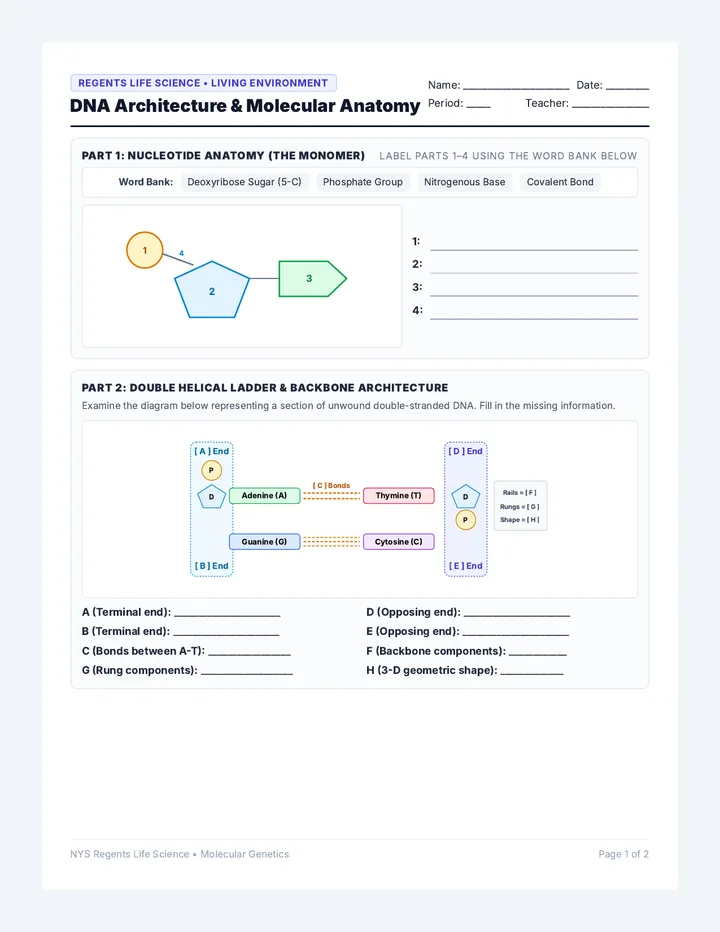
DNA Anatomy Worksheet
Regents Life Science • Living Environment
DNA Architecture & Molecular Anatomy
Name: ______________________ Date: _________
Period: _____ Teacher: ________________
Part 1: Nucleotide Anatomy (The Monomer) Label parts 1–4 using the word bank below
Word Bank: Deoxyribose Sugar (5-C) Phosphate Group Nitrogenous Base Covalent Bond
1 4 2 3
1:
2:
3:
4:
Part 2: Double Helical Ladder & Backbone Architecture
Examine the diagram below representing a section of unwound double-stranded DNA. Fill in the missing information.
[ A ] End [ B ] End P D Adenine (A) [ C ] Bonds Thymine (T) [ D ] End [ E ] End D P Guanine (G) Cytosine (C) Rails = [ F ] Rungs = [ G ] Shape = [ H ]
A (Terminal end): ______________________
D (Opposing end): ______________________
B (Terminal end): ______________________
E (Opposing end): ______________________
C (Bonds between A-T): _________________
F (Backbone components): ____________
G (Rung components): ___________________
H (3-D geometric shape): _____________
NYS Regents Life Science • Molecular Genetics Page 1 of 2
Part 3 & 4: Quantitative Analysis & Regents Practice
DNA Architecture Worksheet
Part 3: Quantitative Base Pairing (Chargaff's Rule)
According to Chargaff's rule, Adenine always pairs with Thymine, and Guanine always pairs with Cytosine. In double-stranded DNA: %A = %T and %G = %C. The sum of all four bases must equal 100%.
Problem 1: Yeast Genome Sample
A sample of yeast DNA contains 31% Adenine (A).
Percentage of Thymine (T): ________ %
Percentage of Guanine (G): ________ %
Percentage of Cytosine (C): ________ %
Problem 2: Bacterial Plasmid
A bacterial gene has 38% Cytosine (C).
Percentage of Guanine (G): ________ %
Percentage of Adenine (A): ________ %
Percentage of Thymine (T): ________ %
Part 4: Regents Examination Analysis
1. Which statement correctly describes the relationship between the components of a DNA molecule?
(1) The sugar-phosphate backbone is held together by weak hydrogen bonds.
(2) Deoxyribose sugars bond directly to both the phosphate group and the nitrogenous base.