Nucleotide Blueprints Presentation NUCLEOTIDE BLUEPRINTS
Unlocking the Building Blocks of DNA
The Molecular Puzzle
Imagine you are a molecular engineer. You have three distinct parts that MUST fit together to form a stable structure.
The Requirements:
One central piece with 5 sides.
One piece containing Phosphorus.
One piece that codes for information.
Phosphate
Sugar
Nitrogenous Base
Anatomy of a Nucleotide
Part 1
Phosphate Group
Contains a phosphorus atom surrounded by four oxygen atoms. This forms the "acidic" part of DNA.
Part 2
Deoxyribose
A 5-carbon sugar. Notice the name "Deoxy" - it's missing one oxygen atom compared to ribose.
Part 3
Nitrogenous Base
The "information carrier." There are four types, categorized by their molecular size.
Purines vs. Pyrimidines
PURINES
2 Double-ring structures
Adenine (A)
Guanine (G)
Mnemonic: "Pure As Gold" (Purine = Adenine, Guanine)
PYRIMIDINES
1 Single-ring structures
Cytosine (C)
Thymine (T)
Mnemonic: "CUT the Pie" (Pyrimidines = Cytosine, Uracil, Thymine)
The Backbone of Life
Nucleotides don't just sit there—they bond to form long strands.
Covalent Bonds
Strong bonds that connect the Phosphate of one nucleotide to the Sugar of the next.
Structural Integrity
This "sugar-phosphate backbone" protects the nitrogenous bases hidden inside.
REPEAT N TIMES
Nucleotide Assembly Worksheet Nucleotide Builder
Lesson 1: Molecular Architecture
Student Name:
Date:
1
Classifying the Bases
Before assembling your nucleotides, categorize the following nitrogenous bases. Use the structural hints provided to determine if they are Purines (2 rings) or Pyrimidines (1 ring).
Purines (Double Ring)
Hint: "Pure As Gold"
Pyrimidines (Single Ring)
Hint: "CUT the Pie"
2
Assembly Zone
Cut out the shapes from the manipulative sheet. Glue them below to form four complete nucleotides . Each nucleotide must contain one sugar, one phosphate, and one base.
Nucleotide 1
Nucleotide 2
Nucleotide 3
Nucleotide 4
3
Critical Analysis
1. To which carbon of the deoxyribose sugar is the nitrogenous base attached?
2. What type of chemical bond connects the phosphate of one nucleotide to the sugar of the next? Why must this bond be strong?
3. Explain the primary difference in structure between a purine and a pyrimidine. How did this affect how you built your models?
Nucleotide Manipulatives Sheet Nucleotide Manipulatives
For use with the Nucleotide Assembly Worksheet. Cut out carefully!
Deoxyribose Sugars
SUGAR
SUGAR
SUGAR
SUGAR
SUGAR
SUGAR
Phosphate Groups
P
P
P
P
P
P
Nitrogenous Bases
ADENINE (A)
GUANINE (G)
ADENINE (A)
GUANINE (G)
CYTOSINE (C)
THYMINE (T)
CYTOSINE (C)
THYMINE (T)
Teacher Note: Print on heavy cardstock if available.
Blueprint of Life // DNA Series
The Chargaff Code Presentation THE CHARGAFF CODE
Cracking the Math of Base Pairing
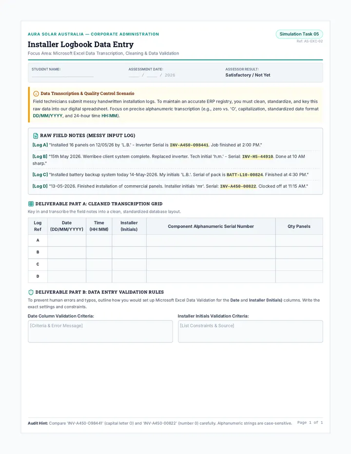
A = T G = C
The Data Mystery
In the late 1940s, Erwin Chargaff noticed something strange about DNA from different species.
"The proportion of adenine is always equal to thymine, and the proportion of guanine is always equal to cytosine."
Why does this matter?
Organism A% T% Human 30.9 29.4 Chicken 28.0 28.4 Yeast 31.3 32.9
≈ 1 : 1 RATIO
Complementary Pairing
A
T
Adenine & Thymine
2 Hydrogen Bonds
G
C
Guanine & Cytosine
3 Hydrogen Bonds
RULE: Purine (Big) + Pyrimidine (Small) = Constant Width
The Power of Hydrogen
Why use Hydrogen Bonds to connect the bases?
Easy to Unzip
Hydrogen bonds are weak enough to be broken easily during DNA replication or transcription.
Specific Pairing
The shape of the bases only allows for specific hydrogen bonding matches (A-T, G-C).
The Challenge
If a DNA sample contains 22% Adenine...
What is the % of Thymine? ??%
What is the % of Cytosine? ??%
Check your Data Detective Worksheet to solve!
Data Detective Worksheet Data Detective
Lesson 2: Analyzing Chargaff's Rules
Scientist:
Date:
The Investigation
In 1950, Erwin Chargaff published a paper that changed how we look at DNA. Below is a simplified data set similar to what he observed. Use your knowledge of Complementary Base Pairing to solve for the missing values.
Part 1: Solving the Puzzle
Organism Adenine (A) Thymine (T) Guanine (G) Cytosine (C) Human 30.9% _______ _______ 19.8% Wheat _______ 27.3% 22.7% _______ Yeast 31.3% _______ 18.7% _______ E. Coli _______ _______ 26.0% _______
Part 2: Evidence Analysis
1. Look at your completed table. What pattern do you see between the percentages of Adenine and Thymine across all species?
2. If a DNA sample had 15% Guanine, explain exactly how you would calculate the percentage of Thymine.
3. Why do you think the percentages for A and T (or G and C) aren't always exactly identical in real-world lab data? List two possible reasons.
The Bonus Challenge
A newly discovered deep-sea virus has a genetic molecule with 20% Adenine, 30% Thymine, 25% Guanine, and 25% Cytosine.
Does this virus follow Chargaff's Rules? Why or why not? What might this suggest about its genetic structure?
LAB REF: DNA-02-CHARGAFF
Confidential Researcher Notes
Data Detective Answer Key Answer Key: Data Detective
Lesson 2 Teacher Resource
Teacher Use Only
Part 1: The Data Table
Organism A % T % G % C % Human 30.9% 30.9% 19.8% 19.8% Wheat 27.3% 27.3% 22.7% 22.7% Yeast 31.3% 31.3% 18.7% 18.7% E. Coli 24.0% 24.0% 26.0% 26.0%
Part 2: Evidence Analysis
1. Pattern between A and T?
Across all species, the percentage of Adenine is nearly identical to the percentage of Thymine. This suggests that they pair together in the DNA structure.
2. Calculation for 15% Guanine?
If G = 15%, then C must also be 15% (Chargaff's Rule). 15% + 15% = 30%. The remaining 70% must be A + T. Since A and T are equal, you divide 70% by 2. T = 35%.
3. Why not exactly identical?
Experimental/measurement error during lab testing.
DNA mutations (though rare, they can slightly alter ratios).
Presence of single-stranded DNA in some viral samples or during replication.
Bonus Challenge Solution:
No, this virus does NOT follow Chargaff's rules (A does not equal T).
This suggests the virus likely has single-stranded DNA (ssDNA) rather than a double helix. Without a second strand to provide complementary pairing, the base ratios can be arbitrary.
Twisted Ladder Presentation TWISTED LADDER MODELS
Constructing the Double Helix
Antiparallel Strands
The two strands of DNA don't run in the same direction. They are like a two-lane highway.
Directionality
One strand runs 5' to 3'
The other strand runs 3' to 5'
5'
3'
3'
5'
The Double Helix
DNA isn't a flat ladder. It twists into a 3D spiral. This shape provides extreme stability and protects the genetic code inside.
Right-Handed
The helix twists to the right in biological DNA (B-DNA).
Major/Minor Grooves
The uneven twist creates gaps where proteins can attach.
10 Base Pairs
There are approximately 10 rungs per full 360° turn.
Building the Blueprint
Today, you are building a physical model. Your model MUST demonstrate:
Antiparallel directionality (5' to 3')
Correct base pairing (A-T, G-C)
The distinct double helix twist
Materials List
• Twizzlers / Pipe Cleaners (Backbone)
• Gummy Bears / Beads (Bases)
• Toothpicks (Hydrogen Bonds)
• Tape / Labels (Directionality)
Visualizing the Twist
Teacher: Insert a short animation of the DNA Double Helix rotating here to emphasize the antiparallel structure.
DNA Modeling Guide Worksheet Twisted Ladder Build
Lesson 3: Double Helix Construction Guide
Modeler:
Date:
Structural Requirements
Antiparallel orientation (5' vs 3')
10 rungs minimum
Correct base pairing colors
360-degree twist demonstrated
Building Supplies
• 2x Pipe Cleaners
• 20x Toothpicks
• Red Beads (A)
• Blue Beads (T)
• Green Beads (G)
• Yellow Beads (C)
Construction Phases
1
Label your Strands
Take two pipe cleaners. Using small pieces of masking tape, label the top of one "5-prime" and the bottom "3-prime". On the second pipe cleaner, label the top "3-prime" and the bottom "5-prime".
2
Prepare the Rungs
Slide two beads onto each toothpick. Remember: Red always goes with Blue (A-T) and Green always goes with Yellow (G-C). Create 10-12 rungs.
3
Assemble the Ladder
Poke the ends of the toothpicks through the pipe cleaners at even intervals. You should now have a flat, rectangular ladder.
4
The Helix Twist
Hold the top of your ladder steady and gently rotate the bottom clockwise to create the double helix spiral.
Structural Reflection
1. Why was it important to flip the direction of the tape labels on the second strand? What does "antiparallel" actually mean in terms of molecular orientation?
2. In your model, what do the toothpicks represent? Why did we use a single toothpick for the bond, rather than gluing the beads directly to each other?
3. Observe your finished helix. Locate a "Major Groove" (a large gap between the twisting backbones) and a "Minor Groove" (a small gap). Why might the size of these gaps matter for biological processes like reading DNA?
Teacher Signature / Photo Spot: Show your stable twisted helix to the instructor for a "Structural Integrity Check."
Strawberry Lab Manual Strawberry Lab
Lesson 4: Macroscopic DNA Extraction
Investigator:
Lab Station:
Safety Protocols
Goggles must be worn at all times. Ethanol is flammable; keep away from heat. Do not consume any lab materials, including the strawberries.
Purpose
To isolate and visualize DNA from octoploid strawberry cells. We will use mechanical force to break cell walls, detergent to dissolve cell membranes, and ethanol to precipitate the DNA molecules.
Checklist
[ ] Strawberry
[ ] Zip-lock Bag
[ ] Lysis Buffer
[ ] Coffee Filter
[ ] Funnel
[ ] Cold Ethanol
[ ] Wooden Skewer
Laboratory Procedure
01
Mechanical Lysis: Place one strawberry in the zip-lock bag. Squeeze out the air and seal it. Mash the strawberry thoroughly for 2 minutes. Do not pop the bag!
02
Chemical Lysis: Open the bag and add 10mL of extraction buffer (soap/salt solution). Reseal and mash gently for another minute to dissolve nuclear membranes.
03
Filtration: Set up a funnel over a test tube with a coffee filter. Pour the strawberry mash into the filter and wait for the liquid (filtrate) to collect in the tube.
04
Precipitation: Tilt the test tube and slowly pour 5mL of ice-cold ethanol down the side. Do not mix! Look for white, cloudy strands forming in the top layer.
Observations & Analysis
Sketch your results
Test Tube View
Describe the appearance of the DNA you extracted:
Why does the DNA become visible when we add ethanol, but not before?
Critical Thinking
We used dish soap in our extraction buffer. Based on what you know about the structure of a cell (specifically the plasma and nuclear membranes), why was the soap necessary to get the DNA out of the cell?
Lab Reflection Exit Ticket Laboratory Debrief
Extraction Exit Ticket
Name:
Section:
1 We built 3D models with beads and pipe cleaners. How did what you saw in the test tube today differ from those models?
2 DNA is soluble (dissolves) in water, but insoluble in alcohol. Which liquid did we use to make the DNA "precipitate" (clump together)?
3 Why can we see the DNA in the test tube with our naked eye, even though a single DNA molecule is microscopic?
Hand to teacher before leaving the lab.
The Race for Photo 51 Presentation THE RACE FOR PHOTO 51
The Untold Discovery of the Double Helix
The Players
James Watson
The Model Builder
Francis Crick
The Physicist
Rosalind Franklin
The Crystallographer
Maurice Wilkins
The Lab Leader
Exhibit A: Photo 51
The "X" Pattern
This isn't a picture of DNA. It's a Diffraction Pattern —a shadow cast by DNA when hit with X-rays.
The "X" shape revealed the molecule was a Helix .
The spacing of the dots showed the Width of the molecule.
Collaboration or Theft?
Maurice Wilkins showed Photo 51 to James Watson without Rosalind Franklin's permission.
The Conflict
Watson and Crick used Franklin's precise measurements to finish their model. They published in 1953 and won the Nobel Prize in 1962. Rosalind Franklin passed away in 1958 and was never officially recognized by the Nobel committee.
The Power of Evidence
Science is rarely a "lone genius" moment. It requires:
Chargaff's Data
The "rules" of pairing (A=T, G=C)
Franklin's Photo
The physical shape (Helix)
Watson/Crick
The 3D synthesis (Modeling)
Discovery Case Study Worksheet Mystery Case File
Lesson 5: The Race for the Double Helix
Investigator:
Date:
The Evidence: Photo 51
In 1952, Rosalind Franklin spent 100 hours of X-ray exposure to capture a single image of DNA. She was an expert in X-ray crystallography—the science of determining molecular structure by measuring how crystals diffract X-rays.
Without her knowledge, her colleague Maurice Wilkins showed the image to James Watson. Upon seeing it, Watson famously said, "My jaw fell open and my pulse began to race."
Specimen: B-DNA
Phase 1: Scientific Interpretation
1. The "X" in the center of the image is the mathematical signature of a helix. If the image had shown a series of parallel lines instead, what might that have suggested about DNA's shape?
2. Franklin’s precise measurements showed that the distance between the two strands was consistent. How does this support Chargaff's Rule (Purine + Pyrimidine)?
Phase 2: Ethics in Discovery
3. Maurice Wilkins shared Franklin's data with Watson and Crick without her permission. Do you believe this was "theft," or simply a necessary collaboration to advance science? Justify your stance.
4. Watson, Crick, and Wilkins won the Nobel Prize in 1962. Franklin had died of cancer (likely caused by X-ray exposure) in 1958. Nobel Prizes are not awarded posthumously (after death). In your opinion, how should history remember Rosalind Franklin?
The Final Verdict
In one sentence, explain how the discovery of DNA structure was a synthesis of data, technology, and modeling.
Case #1953-HELIX
Archival Record // King's College London