Smell Geometry Slides The Geometry of Smell
Molecular Geometry in Biology • Lesson 1
The Mystery Vials
Chemical Facts
Both vials contain the formula C10H14O
Both have the same atomic connections
Both have the same boiling points
So why does Vial A smell like Spearmint and Vial B smell like Caraway Seeds (Rye)?
A
Spearmint
B
Caraway
"One formula, two worlds of experience."
It's All in the Mirror
Meet the Enantiomers
These are molecules that are non-superimposable mirror images of each other.
Think about your hands:
They are mirror images.
They cannot be stacked perfectly on top of each other.
L-Carvone
D-Carvone
The Receptor "Glove"
Structure = Function
1
Molecular Docking
Olfactory receptors are proteins with specific 3D "pockets." Only a molecule with the perfect shape can fit.
2
Signal Induction
When the molecule binds, it changes the protein's shape, triggering a nerve impulse to the brain.
3
Pattern Recognition
The brain interprets which set of receptors were activated as a specific "smell."
"Try putting a right-handed glove on your left hand. It physically doesn't fit correctly. Receptors work the same way."
Why Does Geometry Matter?
Medicine
Most drugs only work if they are the correct "isomer." The wrong shape might be useless or even toxic.
Flavoring
Artificial sweeteners and flavorings must perfectly match the geometry of our taste receptors.
Evolution
Life is "homochiral"—it primarily uses one specific shape of amino acids and sugars.
Next Step: Modeling Your Own Enantiomers!
Molecular Mirror Worksheet Molecular Mirror Lab
Chemistry & Biology • Geometry of Smell
Student Name:
Date:
1
The Sensory Mystery
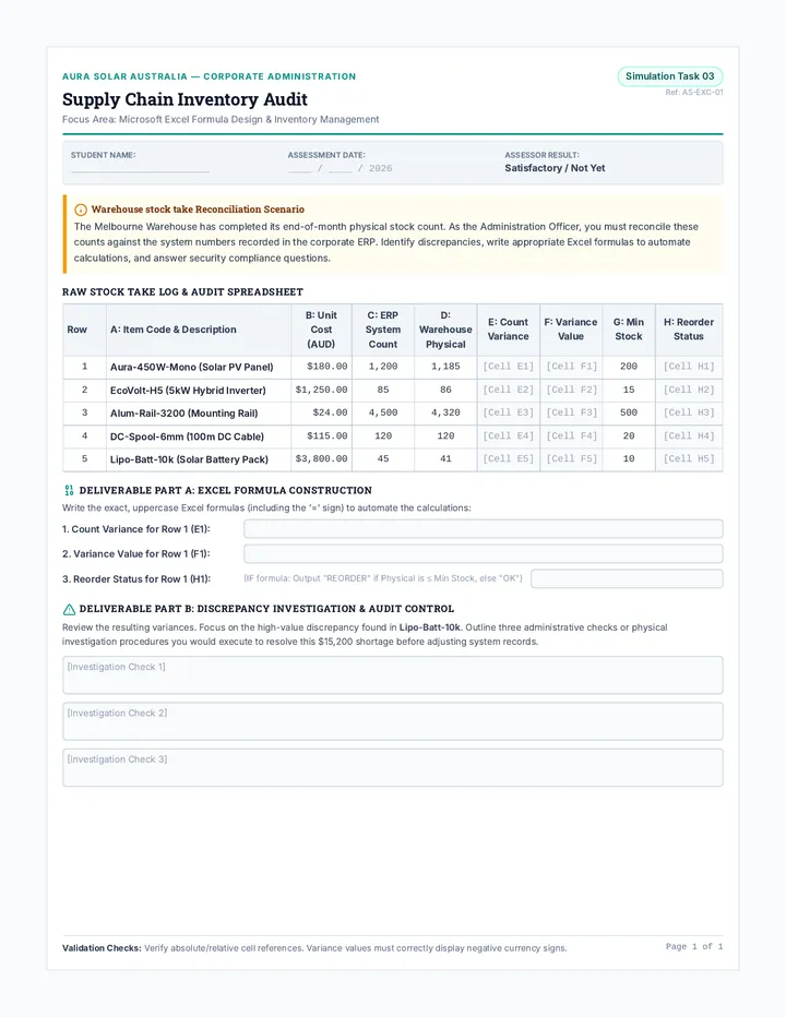
You have been provided with two vials, labeled Sample A and Sample B. Both contain the same chemical formula: C10H14O.
Sample A
Describe the scent:
What does it remind you of?
Sample B
Describe the scent:
What does it remind you of?
Analysis Question
If both molecules are made of the exact same atoms, why do they smell different to your brain?
2
The Hand Analogy
Place your hands palm-down on the table. They are mirror images of each other. Try to slide one on top of the other so all fingers line up.
Terminology Check:
Chiral A molecule that is not identical to its mirror image.
Enantiomer The specific name for a pair of mirrored molecules (like Sample A and B).
3
Molecular Sketching
Below is a simplified 2D drawing of Carvone . To make them 3D enantiomers, we use "wedges" (pointing toward you) and "dashes" (pointing away).
Draw Mirror A Here
L-Carvone (Spearmint)
Draw Mirror B Here
D-Carvone (Caraway)
4
The Receptor Pocket
Think of your olfactory receptor as a right-handed glove . Why can't a "left-handed" molecule activate a "right-handed" receptor effectively? Use the concept of 3D fit in your answer.
End of Lab Activity
Lock and Key Slides Lock and Key Biology
Precision in Every Reaction • Lesson 2
The Bent Key Problem
Imagine your house key has been filed down or slightly bent by just 1 millimeter.
Result: Jammed.
The lock doesn't turn. The door stays closed. The system fails.
In Biology,
Shape is the Code.
Biological Catalysts
Enzymes
Proteins that speed up chemical reactions in our bodies by millions of times .
Substrate
The specific molecule that an enzyme acts upon.
98.6°F
Without enzymes, your body temperature is too cold for life-sustaining reactions to happen fast enough.
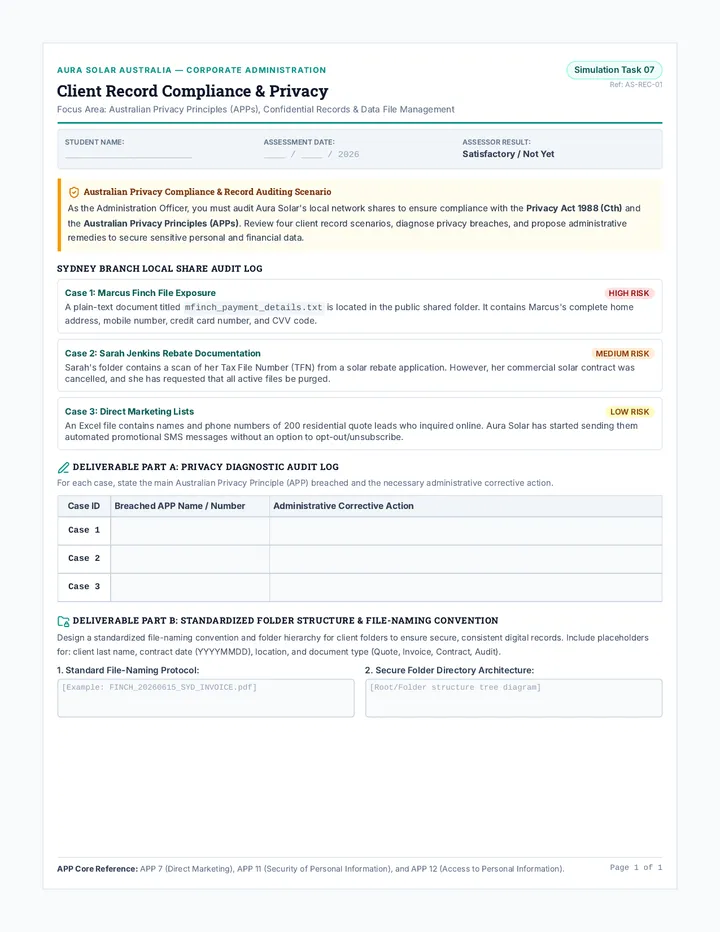
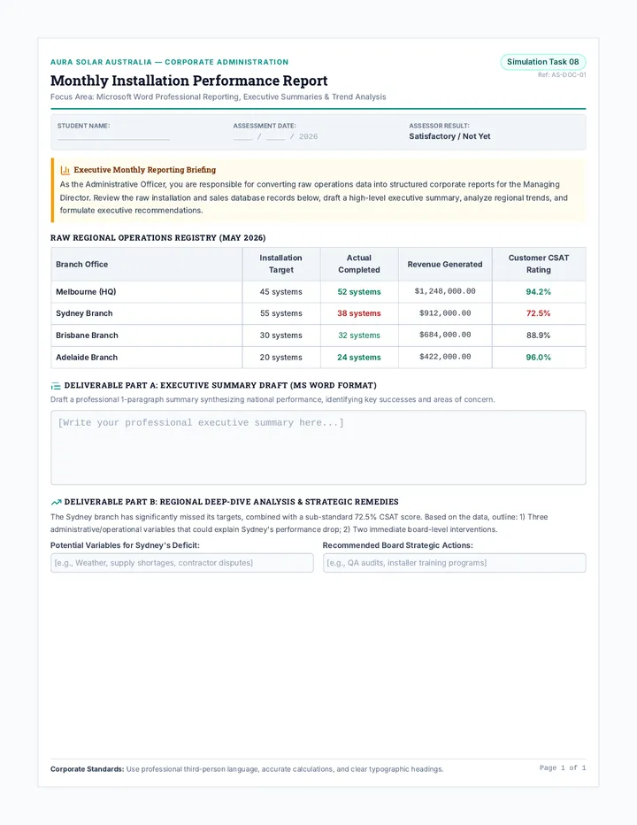
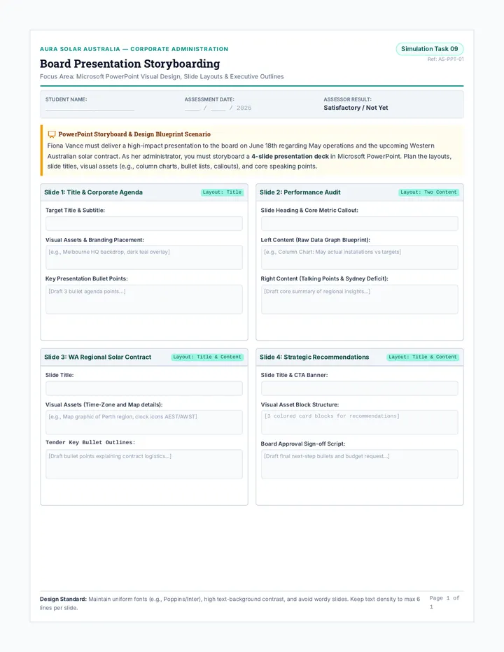
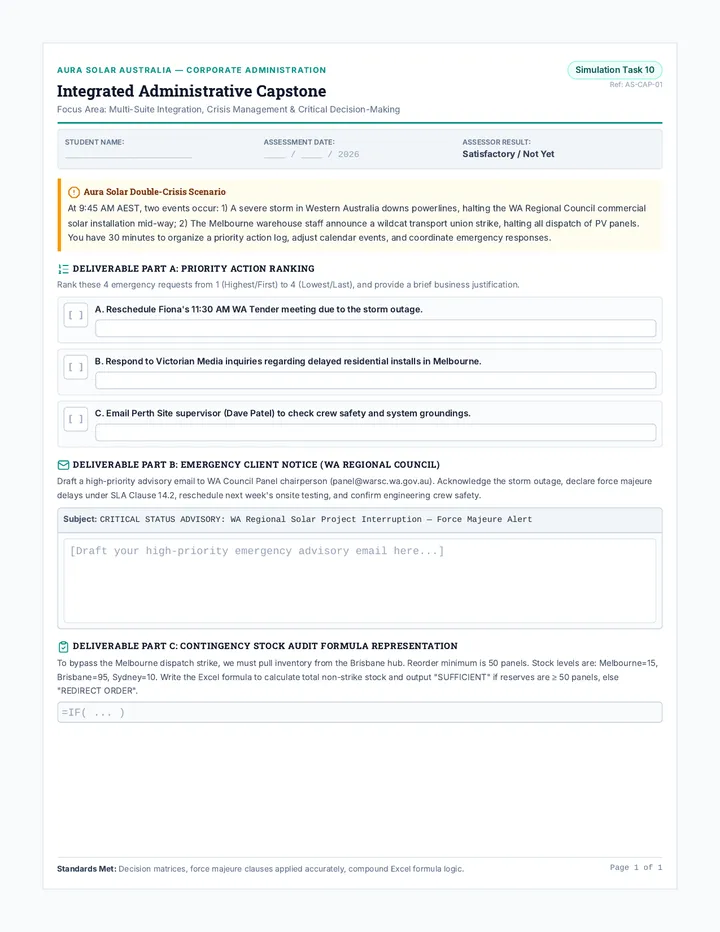
The "Active Site"
Where Geometry Happens
Enzyme
SUBSTRATE
The Active Site is a specific pocket with a unique geometric shape and chemical environment.
Only substrates with the exact complementary shape can fit and bind.
After the reaction, the enzyme is recycled and ready for the next "key."
Why So Picky?
Biological Safety
Specificity prevents "accidental" reactions. If an enzyme could break down anything , it might start breaking down you .
"Imagine a master key that could open every door in the city. Convenient for you, but a disaster for security."
Geometric Check:
Correct Bond Angles?
Correct Surface Area?
Correct Molecular Volume?
Correct Charge Distribution?
Enzyme Precision Simulation Enzyme Precision Lab
Lesson 2 • The Lock and Key Model
Student Name:
Class Period:
A
Mechanical Analysis
Observe the keys and locks provided at your station. For each scenario, predict if the "reaction" (opening the lock) will occur.
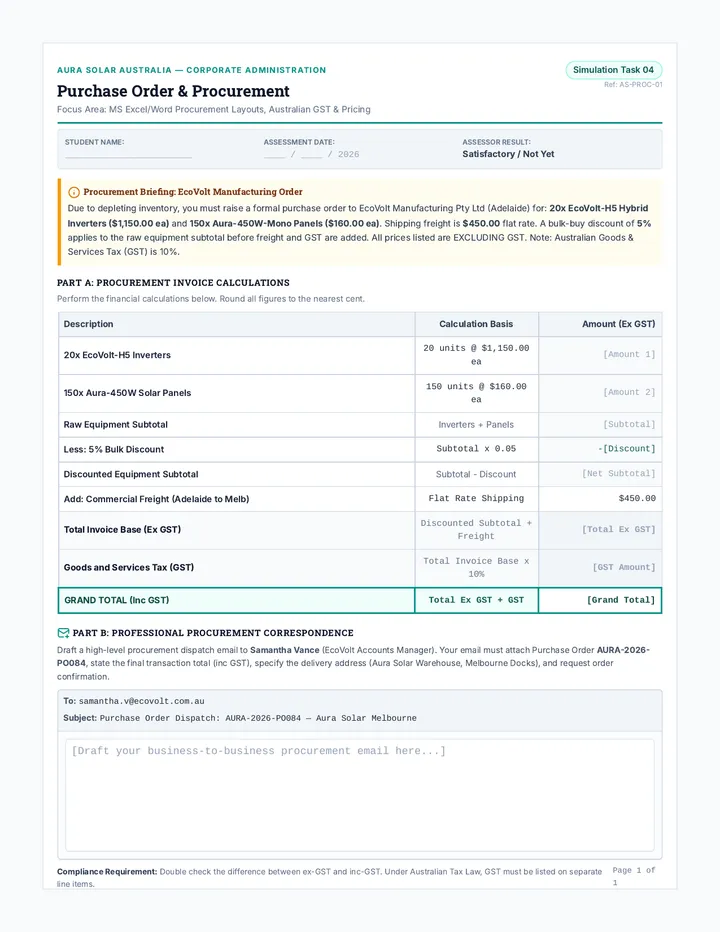
Key Condition Geometric Change Outcome Standard Match No change; exact 3D complement. Bent Tip 5 degree angle shift in key geometry.
|
| Filed Notch | Reduced vertical depth by 0.5mm. |
|
| Tape Obstruction | Introduction of a "blocking" surface. |
|
B
Biological Translation
1. In this simulation, what does the 'Key' represent at a molecular level?
2. What represents the 'Enzyme's Active Site'?
3. Why is it significant that a 0.5mm change prevented the mechanical key from working? Relate this to molecular geometry (bond lengths/angles).
C
Design Challenge
Below are three active sites (pockets). Sketch a substrate molecule for each that would fit perfectly into the geometry shown.
Active Site Alpha
Design for Complementary Fit
Active Site Beta
Design for Complementary Fit
Active Site Gamma
Design for Complementary Fit
Enzyme Kinetics Model v1.2
Molecular Specificity
Mimicry Masterclass Slides Molecular Mimicry
The Chemistry of Imposters • Lesson 3
Why Does Coffee Work?
Your brain produces a molecule called Adenosine throughout the day.
"When Adenosine binds to its receptor, it sends a signal: STAY TIRED ."
Caffeine stops this signal. How?
Adenosine
≈
Caffeine
"The Ultimate Identity Theft"
Chemical Imposters
Agonists
Molecules that mimic the shape AND trigger the response.
"The fake key that actually turns the lock."
Antagonists
Molecules that mimic the shape BUT block the response.
"The fake key that jams the lock so the real one can't get in."
Caffeine is an Adenosine Antagonist!
Designing Cures
Step 1: Map the Receptor
Scientists use X-ray crystallography to see the exact 3D "pocket" of a receptor.
Step 2: Computer Modeling
Virtual molecules are "docked" into the receptor to find the best geometric fit.
Step 3: Synthesis
Chemists build the molecule atom by atom to ensure correct bond angles.
Common Mimics
Aspirin Mimics Prostaglandins
Beta-Blockers Mimics Adrenaline
Antihistamines Mimics Histamine
Morphine Mimics Endorphins
Why do drugs have side effects?
"Because a key that fits your front door might also accidentally fit the neighbor's garage."
In chemistry, mimicry is rarely 100% perfect. A drug designed for the brain might have a similar enough shape to bind to receptors in the stomach or heart, causing unintended reactions.
Next: Investigating the Adenosine Imposter!
Molecular Imposters Case Study The Molecular Imposter
Pharmacology Case Study • Lesson 3
Research Briefing No. 402
Geometry & Signal Blocking
Case Study: The Sleep Signal
Adenosine is a neurotransmitter that tells your heart to slow down and your brain to prepare for sleep. It builds up while you are awake. To work, it must bind to Adenosine Receptors in the brain.
Caffeine is a plant alkaloid that happens to share a remarkably similar geometric profile to the double-ring structure of Adenosine. When you consume caffeine, it travels to the brain and enters the receptor pocket first.
"Structural Similarity: ~85%"
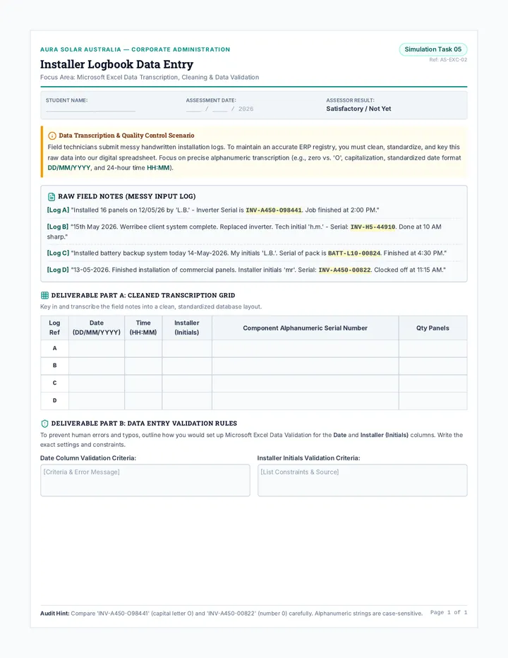
Comparison Data
Mechanism of Adenosine:
Mechanism of Caffeine:
Analysis: The Blocker Effect
Caffeine binds to the receptor but does NOT trigger the "Sleep Signal." Instead, it just sits in the pocket, preventing Adenosine from entering.
1. Is caffeine an Agonist or an Antagonist? Justify your choice based on the mechanism above.
Explain the functional difference here...
2. Draw a simple conceptual diagram showing an Adenosine Receptor with caffeine blocking the "mouth" of the pocket.
Drawing Area: Show Receptor + Blocker
Future Design
If a patient has chronic insomnia (can't sleep), would a pharmaceutical designer want to create an adenosine agonist or an adenosine antagonist ? Explain your reasoning using the lock and key model.
Student Initials:
Chemistry of Mimicry Project
Protein Shifting Slides Shape Shifters
Protein Folding & Failure • Lesson 4
The Incredible Shrinking Protein
Take a raw egg white: it's clear, liquid, and runny.
Add heat , and it becomes white, solid, and firm.
The Big Question:
"What physically changed in the molecules to turn a liquid into a solid?"
Energy Input
When you add heat, you are vibrating the molecular bonds until they break.
The Origami of Life
1°
Sequence
The chain of amino acids (The string).
2°
Local Shape
Helixes and pleats (The loops).
3°
Final 3D Geometry
The "Active" Form
This is where the Lock and Key geometry comes from.
Proteins are held in their final 3D shape by weak attractions: Hydrogen Bonds , Disulfide Bridges , and Hydrophobic Interactions .
Denaturation
Molecular Unfolding
Denaturation is the process where a protein loses its 3D shape due to external stress.
Temperature
Breaks Hydrogen Bonds
pH Levels
Disrupts Ionic Charges
NO SHAPE = NO FUNCTION
An unfolded enzyme cannot bind its substrate.
Why It Matters
Fevers
A high fever (105°F+) is dangerous because your brain's critical enzymes can start to denature . If they lose their shape, your neurons stop firing.
Medicine
Insulin and other protein drugs must be refrigerated . If they get too warm, the protein unfolds and the medicine becomes useless water.
Next: Lab Activity - The Denaturation Test
The Unfolding Lab Report The Unfolding Lab
Lesson 4 • Protein Denaturation Study
Researcher:
Lab Station:
Experimental Background
Proteins are only functional when folded into a specific three-dimensional geometry. In this lab, we will use egg white (Albumin protein) as a model to observe how molecular shape is disrupted by environmental stressors.
1
Albumin State: Native (Folded)
Visual Appearance (Color/Transparency):
Physical State (Viscosity/Texture):
2
Environmental Stressors
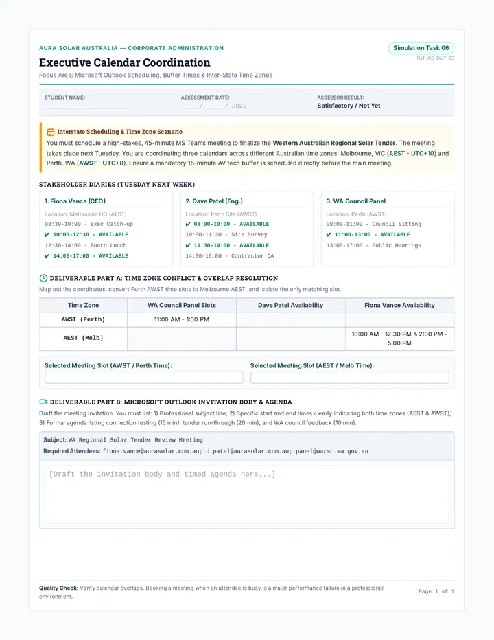
Variable Observation (What changed?) Molecular Explanation Heat (Boiling Water)
| | |
|
Acid (Vinegar/HCl)
| | |
|
Alcohol (Ethanol)
| | |
3
Conclusion
1. Why is denaturation generally irreversible (i.e., you can't "un-cook" an egg)?
2. In terms of "Lock and Key" biology, what happens to the enzyme's Active Site during denaturation?
Thermodynamic Stability Index • Albumin Study
The Blocker Challenge Slides Classified Mission
The Blocker Challenge
Molecular Defense Strategy • Lesson 5
Threat Assessment
A new theoretical pathogen, the "Alpha-Z Virus," infects human cells by binding to a specific protein receptor on the cell surface.
Your Mission:
Design a synthetic antagonist molecule that fits the receptor better than the virus does, effectively blocking the infection.
Pathogen: ALPHA-Z
Geometric Match: HIGH
Design Constraints
1. Geometry
The molecule must perfectly complement the 3D "pocket" of the Alpha-Z receptor.
2. Polarity
Charge distribution must match. Positive areas of the receptor need negative areas on your molecule.
3. Stability
Your design must use stable covalent bonds that won't denature at body temperature (37°C).
Target: Receptor-ZX
Binding Pocket Beta-9
Pocket Profile
Volume: 450 cubic angstroms
Interior Charge: Highly Positive
Shape: Cylindrical Base
Engineer Note:
"If the receptor interior is positive, what functional groups will you add to your molecule to ensure it 'sticks'?"
Engineers to Stations
Use your Blocker Design Portfolio to draft your molecule. You must provide a 3D sketch, a list of atoms used, and a justification for why your geometry works.
Time: 45 Mins
Teams: Pairs
The Blocker Challenge Project Portfolio Design Div.
Blocker Design Portfolio
Operation: Alpha-Z Containment • Final Assessment
Clearance Level 4
Lead Engineer:
Sector ID:
Mission Intelligence
The Alpha-Z Virus binds to Receptor-ZX , a protein located on the surface of human lung cells. The virus fits into a "binding pocket" that is primarily positively charged and shaped like a deep, rounded cylinder. Your task is to design a synthetic molecule (an antagonist ) that fits this pocket perfectly to block the virus.
01
Geometric Blueprint
Primary Design Workspace: Draw 3D Molecule
Chemical Composition
Functional Groups Used:
Atom Inventory:
Meets Volume Limit
Remember to use 'wedges' and 'dashes' to indicate 3D depth in your drawing.
02
Engineering Justification
Geometric Complementarity
How does the 3D shape of your molecule ensure a tighter fit than the Alpha-Z virus?
Charge/Polarity Match
The receptor is POSITIVELY charged. What elements or groups did you use to create attraction?
The Antagonist Argument
Explain why your molecule is a better antagonist than a natural agonist. Why won't your molecule accidentally trigger the cell's internal signal while blocking the virus?
Identity Verified • Bio-Security Protocol Active
Project Alpha-Z Complete